The left main coronary artery supplies approximately 70–80% of the left ventricle’s blood supply. Disease of the unprotected left main — where no bypass graft is present as a safety net — is among the highest-stakes scenarios in interventional cardiology. Left main PCI in the right patient, planned and executed with precision, is a safe and durable alternative to bypass surgery. Dr. Arun Kalyanasundaram performs unprotected left main PCI as a core part of his complex coronary practice.
Technical Success
Mandatory
Pre-procedure Assessment
Evidence Base
The left main coronary artery (LMCA) is typically 1–2 centimetres in length, originating from the left side of the aorta and bifurcating into the left anterior descending artery (LAD) — supplying the front wall of the left ventricle — and the left circumflex artery (LCx) — supplying the lateral and posterior walls. Together, these two arteries supply the majority of the heart’s pumping muscle.
When significant disease develops in the left main — typically defined as stenosis of 50% or greater, or a fractional flow reserve (FFR) below 0.80 — the consequence of untreated obstruction or a complication during intervention is disproportionately severe. Any significant haemodynamic disturbance affecting left main flow can result in rapid haemodynamic collapse.
For these reasons, left main PCI requires more thorough pre-procedural planning, more rigorous intra-procedural monitoring, a higher threshold for haemodynamic support, and a more demanding stenting technique than standard coronary intervention. The gap between the most experienced left main operators and average ones is wider here than in any other coronary territory.
Left main PCI without all four of these components is not contemporary practice. Each is non-negotiable in Dr. Arun’s protocol — applied to every case regardless of anatomical simplicity.
FFR or iFR assessment
Mandatory in intermediate lesions (40–60% stenosis). Angiographic severity alone notoriously overestimates or underestimates the haemodynamic significance of left main disease. FFR ≤ 0.80 or iFR ≤ 0.89 defines functionally significant left main stenosis requiring revascularisation. Without physiological assessment, intervention may be performed on lesions that don’t warrant it — or withheld from lesions that critically do.
IVUS-guided stent sizing
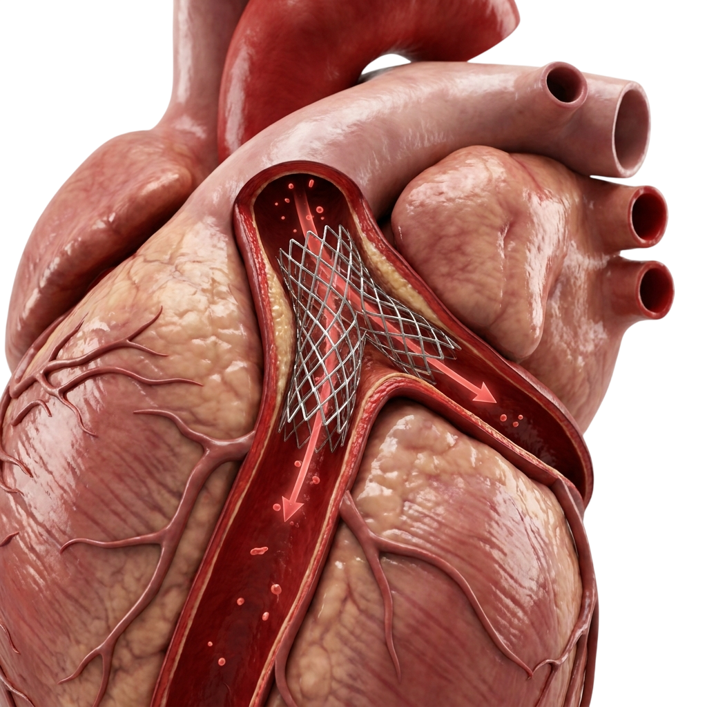
The left main is typically 4–5 mm in diameter — accurate sizing is critical for stent durability. IVUS provides real-time cross-sectional measurement and confirms stent expansion and apposition at bifurcation landing zones in both the left main trunk and the bifurcation branches. Minimum stent area (MSA) targets of 8–10 mm² in the left main have been associated with reduced major adverse cardiac events.
Bifurcation management strategy
Most left main lesions involve the bifurcation. Strategy — provisional single stent vs planned two-stent technique (DK Crush, Culotte, T-and-protrude) — is determined by Medina classification of which segments are diseased and by the SYNTAX subscore. The choice of strategy is made before entering the catheterisation laboratory — never improvised mid-procedure.
Haemodynamic support readiness
For impaired LV function: pre-procedural IABP or Impella placed before the procedure begins — not as rescue after deterioration. Particularly important for ostial or mid-shaft left main lesions where guide catheter engagement itself can transiently obstruct flow. ECMO standby for the most haemodynamically fragile cases.
For patients with low to intermediate anatomical complexity (SYNTAX score below 32–33), contemporary evidence from the EXCEL and NOBLE trials supports left main PCI as a reasonable alternative with comparable five-year outcomes. For patients with more complex anatomy or associated triple-vessel disease, bypass surgery has a durable advantage at longer follow-up. The right choice depends on your specific anatomy, LV function, surgical risk, and preferences — all of which require a multidisciplinary discussion.
Yes. Patients declined for bypass due to high surgical risk — severe lung disease, previous open-heart surgery, frailty, or multiple serious illnesses — can in many cases be managed with left main PCI using a comprehensive haemodynamic support approach. A second opinion from Dr. Arun Kalyanasundaram can assess whether left main PCI is feasible and safe for your specific anatomy.
Most patients stay in hospital for two to three nights, particularly if haemodynamic support was used. Dual antiplatelet therapy (aspirin plus clopidogrel or ticagrelor) is prescribed for at least 12 months. Repeat clinical assessment and functional testing at 3–6 months is standard to confirm adequacy of revascularisation and stent patency.
The SYNTAX score is an angiographic tool that quantifies the anatomical complexity of coronary artery disease. It guides the decision between PCI and bypass surgery for left main disease. ESC guidelines give PCI a Class I indication for SYNTAX score under 22, Class IIa for 23–32, and Class III for over 32 — where bypass is preferred. The score considers lesion location, calcification, tortuosity, bifurcation involvement, and total occlusions across all coronary segments.
Intravascular ultrasound (IVUS) is mandatory in left main PCI because angiography alone is unreliable for vessel sizing in this critical anatomy. The left main is typically 4–5 mm in diameter, and accurate sizing determines stent durability, expansion, and apposition. IVUS provides real-time cross-sectional imaging confirming stent landing zones in both the trunk and bifurcation branches. IVUS-guided left main PCI has been shown to reduce major adverse cardiac events compared with angiography alone.
The left main bifurcation is where the left main coronary artery divides into the left anterior descending (LAD) artery and the left circumflex (LCx) artery. Most left main lesions involve this bifurcation. Stenting strategy — provisional single stent versus planned two-stent technique (DK Crush, Culotte, T-and-protrude) — is determined by the Medina classification of which segments are diseased and by the SYNTAX subscore. Bifurcation involvement increases procedural complexity and is one of the strongest predictors of restenosis.
A typical left main PCI takes 1.5–3 hours depending on bifurcation involvement, calcium burden, and whether two-stent technique is required. Cases requiring calcium modification with rotational atherectomy or intravascular lithotripsy add additional time. Pre-procedural IVUS and FFR/iFR assessment add 15–30 minutes but are essential for planning.
+91 94807 94807
director@ctomd.com
India’s leading CTO PCI specialist.
Cleveland Clinic trained.
Asia-Pacific CTO Club India Director.
Dr. Arun Kalyanasundaram is a Chennai-based CTO PCI specialist providing advanced coronary intervention, CTO angioplasty, blocked artery treatment, second opinions, and treatment planning for patients from Mumbai, Delhi, Bangalore, Hyderabad, Pune, Kolkata, Ahmedabad, Chandigarh, Kochi, Visakhapatnam, and throughout India.
Promed Hospital
1/10A East Coast Road, Kottivakkam
Chennai, Tamil Nadu 600041